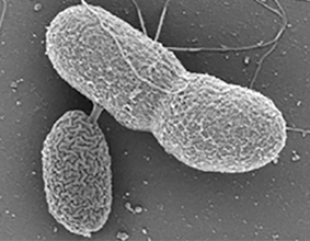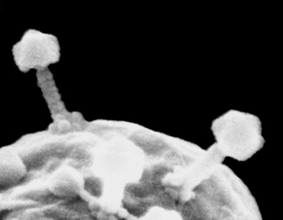
In recent years our research group has developed improved methods for the preservation of biological structures and for delicate, grain-less coating by iridium, for the purpose of high resolution imaging by scanning electron microscopy (SEM). These methods have proven instrumental in direct surface imaging of the nucleus and in revealing the fine architectural features of NPCs. Some of these methods have also turned out to be very useful for imaging a variety of other biological structures in different collaborative projects. We have successfully used SEM imaging for studying fungi and bacteria (Hover et al., 2016; Shemesh et al., 2017) and even myoviruses that infect cyanobacteria in the oceans (Sabehi et al., Proc. Natl. Acad. Sci. USA, 2012). Some of our on-going collaborations focus on yeast chromatin, bacterial injection systems, regenerating ascidians and bacterial communication in an insect’s gut. Other mini-projects are purely curiosity driven expeditions and there’s always something new around the corner…

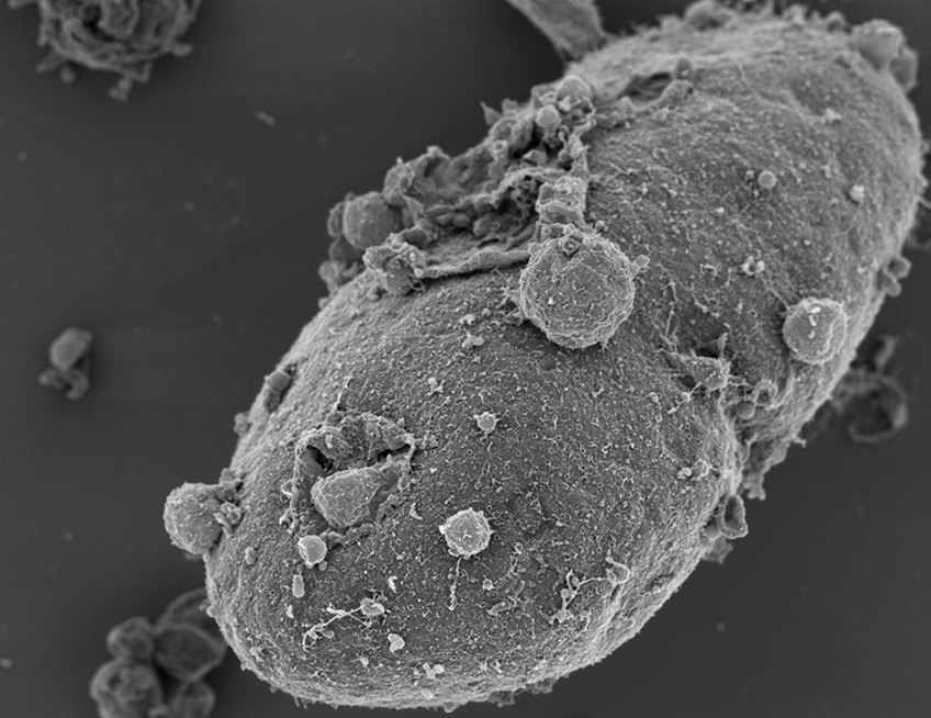
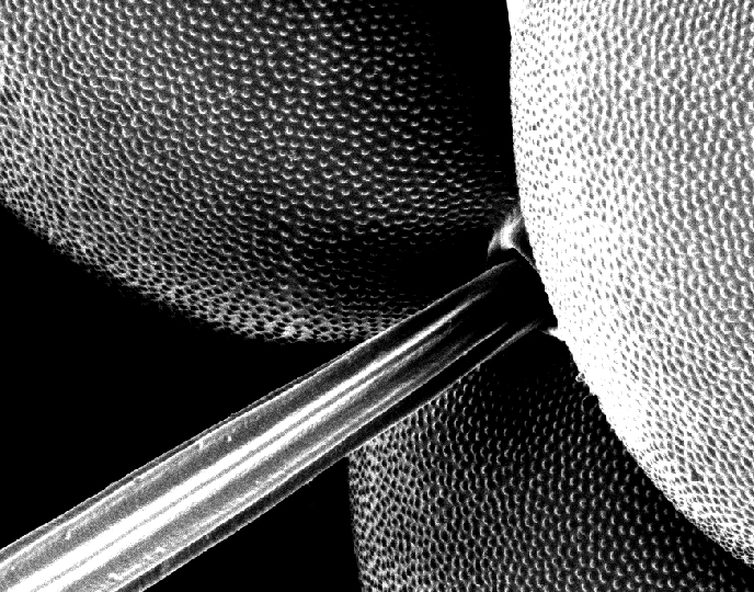
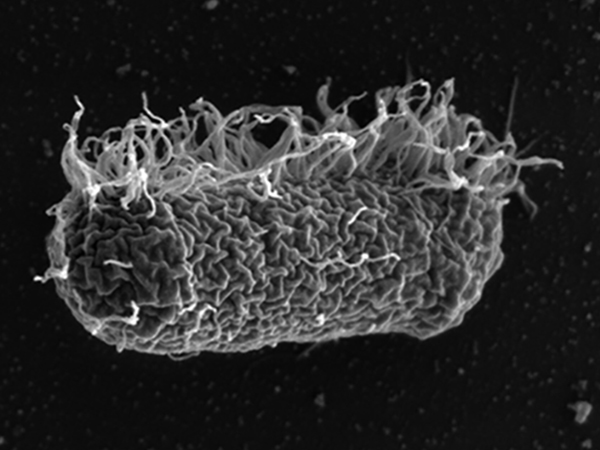
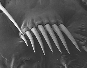
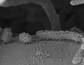
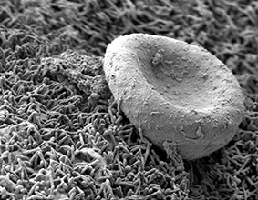
Exposed Nucleus (patient derived fibroblast)Fly Eye (detail) Bad Hair Day Bacterium Bacterial Communication in Fly Gut Minute Pirate Bug Bacterial Attack on Fungal Hyphae Red Blood Cell in Human Placenta Myoviruses